What Is an Animal Cell? Parts, Diagram, and Functions
Every living creature you have ever seen — from the tiny ant crawling across a picnic blanket to the blue whale gliding through ocean depths — shares one fundamental truth: all animals are built from cells. The animal cell is the basic structural and functional unit of animal life, a microscopic factory humming with activity twenty-four hours a day. Understanding how these cells work is not just a biology lesson; it is a window into what makes life itself possible.
What Is an Animal Cell?
An animal cell is a type of eukaryotic cell, which means it contains a true, membrane-bound nucleus that houses its genetic material. Unlike plant cells, animal cells lack a rigid cell wall and chloroplasts, giving them a flexibility and structural simplicity that suits the diverse shapes and functions found throughout the animal kingdom. Every tissue in your body — muscle, nerve, skin, blood — is composed of animal cells specialized for particular tasks, yet all of them share the same core set of internal structures.
What makes animal cells truly remarkable is their ability to cooperate. A single human body contains roughly thirty trillion cells, each one communicating, responding, and adapting to the needs of the whole. When you feel hungry, heal a cut, or fight off a cold, you are witnessing the coordinated efforts of countless individual animal cells working in concert.
A Closer Look at Animal Cell Structure
Think of an animal cell as a bustling city contained within a flexible boundary. Each component, or organelle, has a specific job, and the smooth operation of the entire cell depends on every part doing its work reliably. Let us tour the major structures one by one.
The Cell Membrane: The Gatekeeper
Surrounding every animal cell is the cell membrane, also called the plasma membrane. This is not a simple passive wrapper — it is a sophisticated, selectively permeable barrier made from a phospholipid bilayer studded with proteins. Imagine a soap bubble that can actively decide what enters and what leaves. The membrane allows nutrients like glucose to flow in, waste products to exit, and keeps harmful substances at bay. Receptor proteins embedded in the membrane also act as communication antennae, picking up chemical signals from other cells and the surrounding environment.
The fluid nature of the membrane — often described by the fluid mosaic model — means its components can move laterally, repair small breaches, and even pinch off to form tiny transport vesicles that carry materials deeper into or out of the cell.
The Nucleus: The Command Center
At the heart of the animal cell lies the nucleus, the largest and most visible organelle. Protected by a double-layered nuclear envelope dotted with pore complexes, the nucleus safeguards the cell’s entire genetic blueprint in the form of DNA. During most of the cell’s life, this DNA exists as a diffuse, thread-like material called chromatin. When the cell prepares to divide, the chromatin condenses into the familiar X-shaped chromosomes visible under a microscope.
Inside the nucleus, a dense spherical region called the nucleolus acts as the ribosome factory. Here, ribosomal RNA is synthesized and combined with proteins to form the beginnings of ribosomes, which then exit through nuclear pores to carry out their protein-building duties in the cytoplasm.
Cytoplasm and Cytosol: The Cellular Medium
Filling the space between the cell membrane and the nucleus is the cytoplasm, a gel-like substance composed mostly of water, dissolved salts, and organic molecules. The liquid portion of the cytoplasm is called the cytosol, and it serves as the medium through which organelles, proteins, and other molecules move and interact. Far from being a stagnant pool, the cytoplasm is a dynamic environment where countless biochemical reactions occur every second.
Mitochondria: The Powerhouses
If the nucleus is the brain of the cell, mitochondria are its power plants. These bean-shaped, double-membraned organelles convert energy stored in food molecules into adenosine triphosphate, or ATP — the universal energy currency of the cell. The inner membrane of a mitochondrion is highly folded into structures called cristae, which maximize the surface area available for energy production through cellular respiration.
Mitochondria are unique among organelles because they contain their own DNA and ribosomes, strongly supporting the theory that they originated from ancient bacteria that entered into a symbiotic relationship with early eukaryotic cells billions of years ago. Your mitochondria, passed down exclusively from your mother, are a living link to this deep evolutionary history.
Ribosomes: The Protein Builders
Scattered throughout the cytoplasm and dotting the surface of the rough endoplasmic reticulum, ribosomes are tiny molecular machines constructed from RNA and proteins. Their sole job is to translate genetic instructions carried by messenger RNA into polypeptide chains that fold into functional proteins. Despite their small size, ribosomes work with astonishing speed and accuracy. A single ribosome can add multiple amino acids to a growing protein chain every second, and a typical animal cell contains millions of them.
The Endomembrane System
Several organelles work together in a coordinated network known as the endomembrane system, responsible for synthesizing, modifying, packaging, and transporting proteins and lipids.
The rough endoplasmic reticulum, or rough ER, is studded with ribosomes and specializes in producing proteins destined for secretion or for use in the cell membrane. As these proteins are synthesized, they enter the interior of the rough ER, where they begin folding into their correct three-dimensional shapes.
The smooth endoplasmic reticulum, or smooth ER, lacks ribosomes and focuses on lipid synthesis, detoxification of drugs and poisons, and calcium ion storage. Liver cells, which handle significant detoxification duties, are particularly rich in smooth ER.
The Golgi apparatus functions as the cellular post office. It receives proteins and lipids from the ER, modifies them — often by adding carbohydrate or phosphate tags — sorts them, and packages them into vesicles addressed to specific destinations, whether that is the cell membrane, a lysosome, or the extracellular space.
Lysosomes and Peroxisomes: The Cleanup Crew
Lysosomes are spherical, membrane-bound sacs filled with powerful digestive enzymes capable of breaking down proteins, nucleic acids, lipids, and carbohydrates. They serve as the cell’s recycling and waste disposal system, digesting worn-out organelles, engulfed food particles, and invading bacteria. The interior of a lysosome is maintained at an acidic pH, which optimizes enzyme activity while protecting the rest of the cell should a lysosome rupture.
Peroxisomes perform a related but distinct role, primarily breaking down fatty acids and neutralizing hydrogen peroxide — a toxic byproduct of metabolism — by converting it into water and oxygen using the enzyme catalase.
The Cytoskeleton: Internal Scaffolding
Giving the animal cell its shape, strength, and ability to move is the cytoskeleton, a complex network of three types of protein filaments. Microfilaments, the thinnest, are built from actin and play critical roles in muscle contraction, cell division, and amoeboid movement. Intermediate filaments provide mechanical strength and anchor organelles in place. Microtubules, the thickest filaments, are hollow tubes made of tubulin that serve as tracks for intracellular transport and form the spindle apparatus that separates chromosomes during cell division.
Centrioles, paired cylindrical structures composed of microtubules, organize the spindle fibers during mitosis and also serve as the basal bodies for cilia and flagella — hair-like and whip-like extensions that some animal cells use for movement.
Animal Cell Diagram: Visualizing the Whole
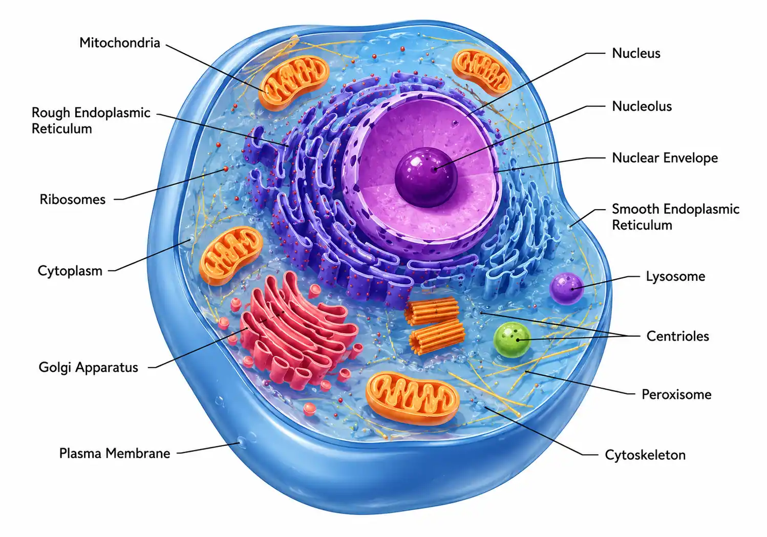
Picture a rounded, irregular shape bounded by a thin outer line — that is the cell membrane. Inside, a large central sphere represents the nucleus, with a smaller dark dot inside it marking the nucleolus. Scattered throughout the interior are numerous oval mitochondria with wavy internal lines, networks of folded membranes representing the rough and smooth ER, and a stack of flattened sacs near the nucleus depicting the Golgi apparatus. Tiny dots sprinkled throughout the cytoplasm and clinging to the rough ER represent ribosomes. Small circles at various locations stand for lysosomes, peroxisomes, and transport vesicles. Thread-like lines crisscrossing the entire interior form the cytoskeleton. This mental image captures the organized complexity of a typical animal cell.
Why Animal Cells Matter
Beyond the diagrams and textbook definitions, animal cells are the reason you can think, move, grow, and heal. Cancer, neurodegenerative diseases, infections, and countless other medical conditions fundamentally arise from malfunctions at the cellular level. Modern medicine, from targeted cancer therapies to gene editing technologies like CRISPR, works by understanding and intervening at the scale of the individual animal cell.
In research laboratories around the world, scientists culture animal cells to test new drugs, study genetic disorders, and explore the fundamental mechanisms of life. Stem cells — unspecialized animal cells with the potential to develop into many different cell types — hold immense promise for regenerative medicine, offering hope for treating conditions ranging from spinal cord injuries to heart disease.
The more we learn about these microscopic building blocks, the more we appreciate the intricate, beautiful, and resilient nature of life itself. Every breath you take, every thought you form, every beat of your heart traces back to the coordinated activity of animal cells — tiny universes operating with breathtaking precision.
Author Bio: Dr. Naomi Chen is a molecular biologist and science educator who translates complex biological concepts into clear, engaging content for curious learners.